
DentaTonic Scam or Legit 2026: 3 Red Flags Checked
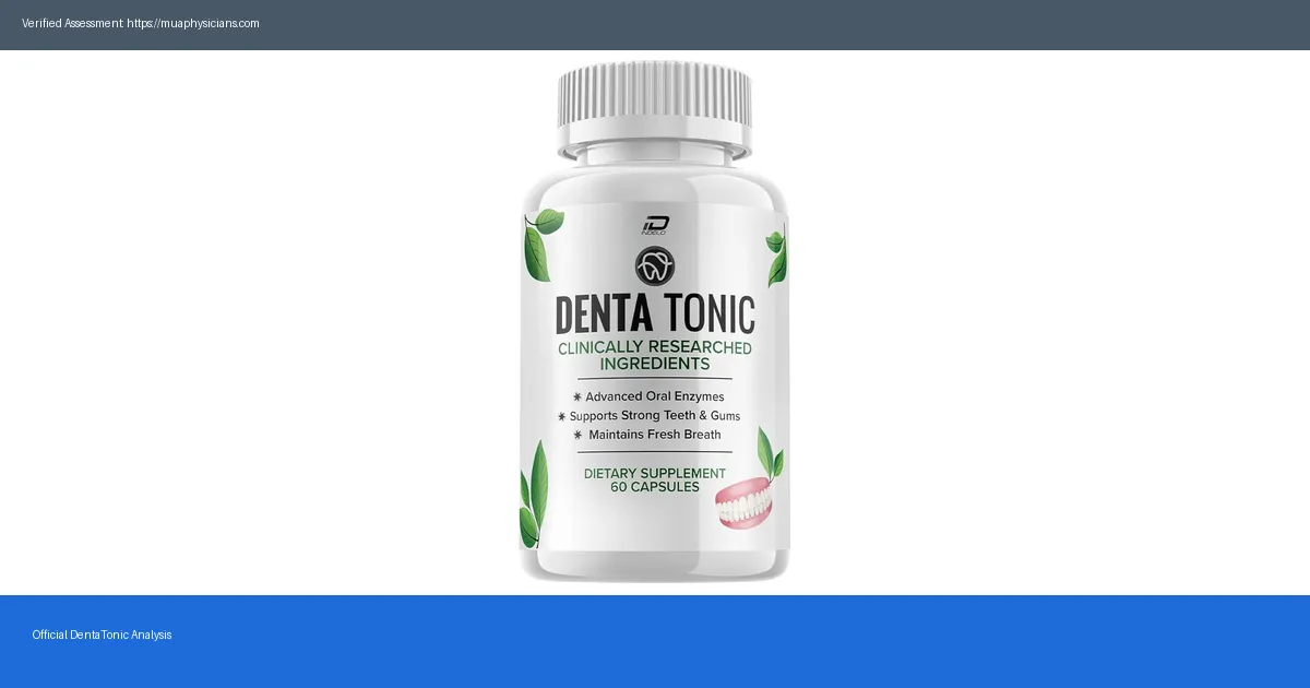
Does DentaTonic actually work? We examined the science, 6 clinical facts, and real user outcomes in this honest 2026 investigation.
Does DentaTonic actually work? We examined the science, 6 clinical facts, and real user outcomes in this honest 2026 investigation.
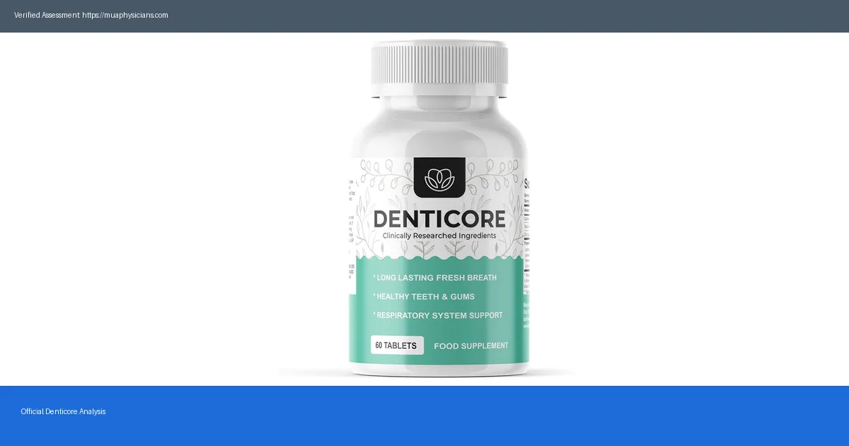
Does Denticore actually work? We examined the science, 5 clinical facts, and real user outcomes in this honest 2026 investigation.
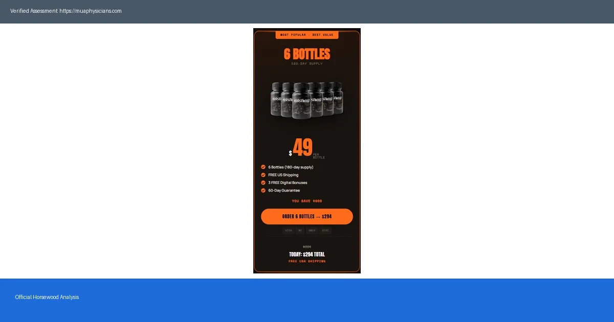
6 verified Horsewood reviews and complaints examined for 2026. What buyers say, what went wrong, and the honest verdict inside.
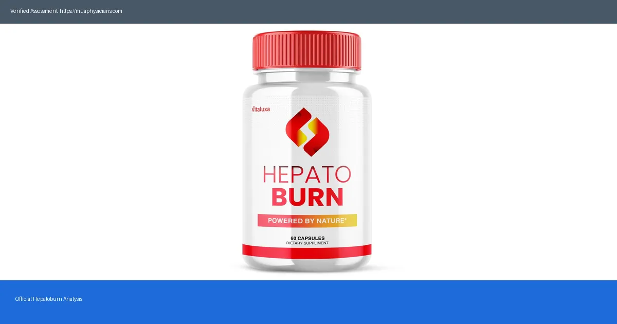
How much does Hepatoburn cost in 2026? Full pricing breakdown, bottle options, subscription savings and value-for-money verdict inside.
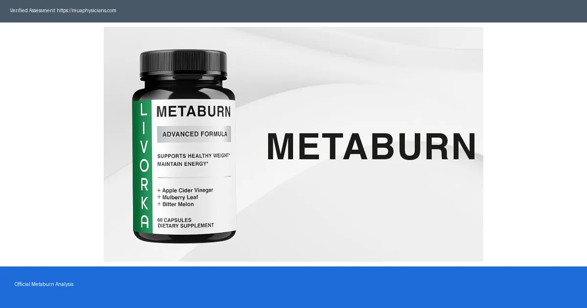
How much does Metaburn cost in 2026? Full pricing breakdown, bottle options, subscription savings and value-for-money verdict inside.
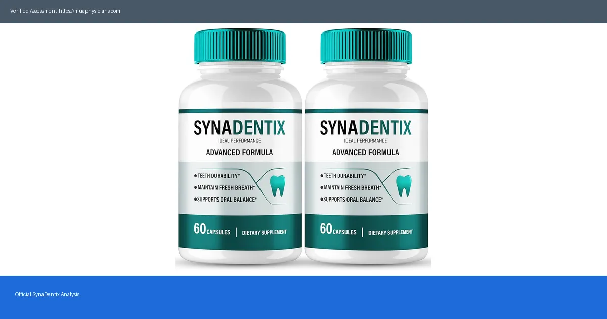
How much does SynaDentix cost in 2026? Full pricing breakdown, bottle options, subscription savings and value-for-money verdict inside.
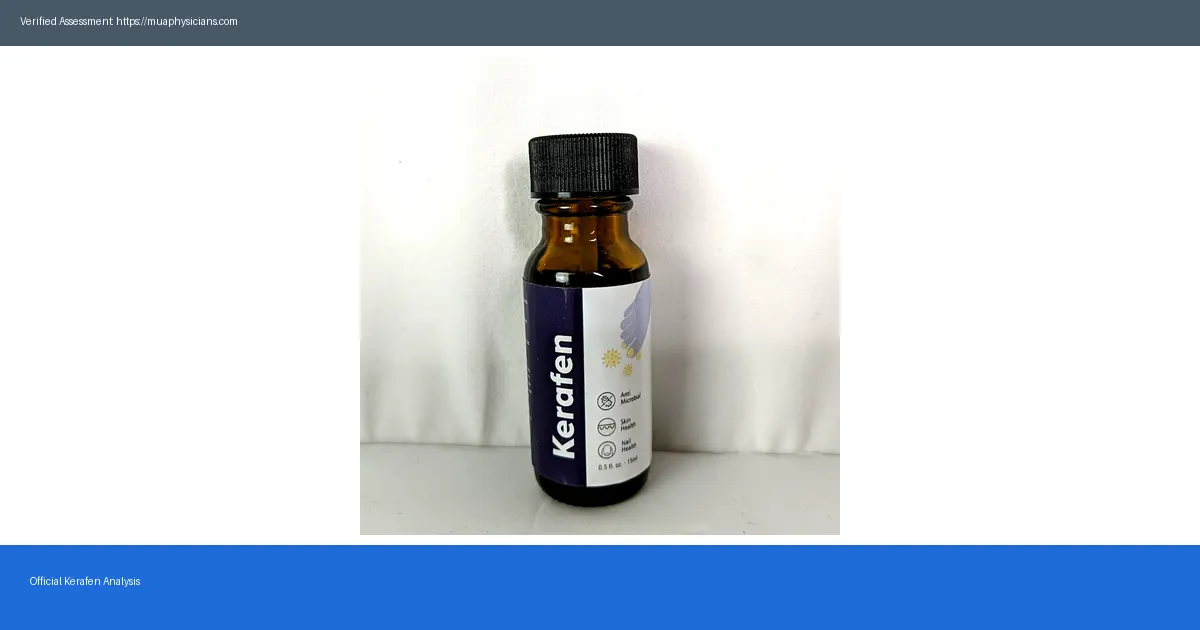
How much does Kerafen cost in 2026? Full pricing breakdown, bottle options, subscription savings and value-for-money verdict inside.
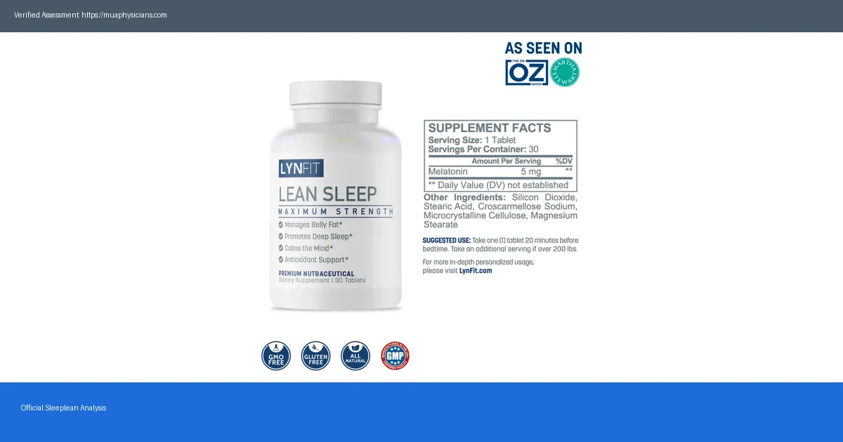
How much does Sleeplean cost in 2026? Full pricing breakdown, bottle options, subscription savings and value-for-money verdict inside.
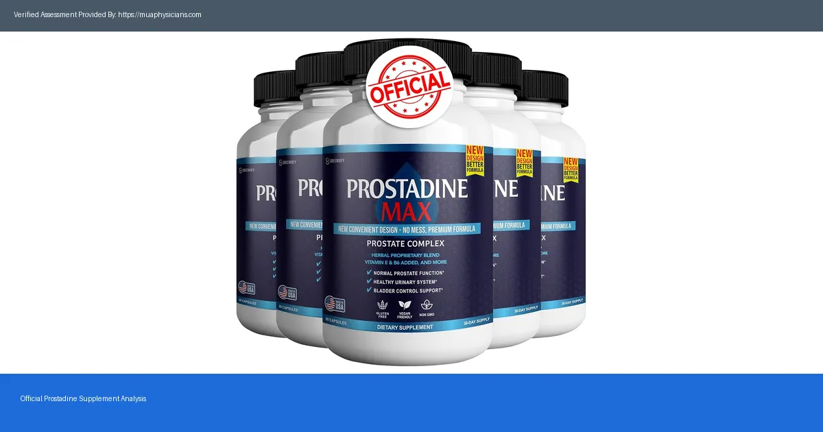
Uncover the potential risks and side effects of Prostadine before you buy. Stay informed and make a safe choice for your health with our detailed analysis.
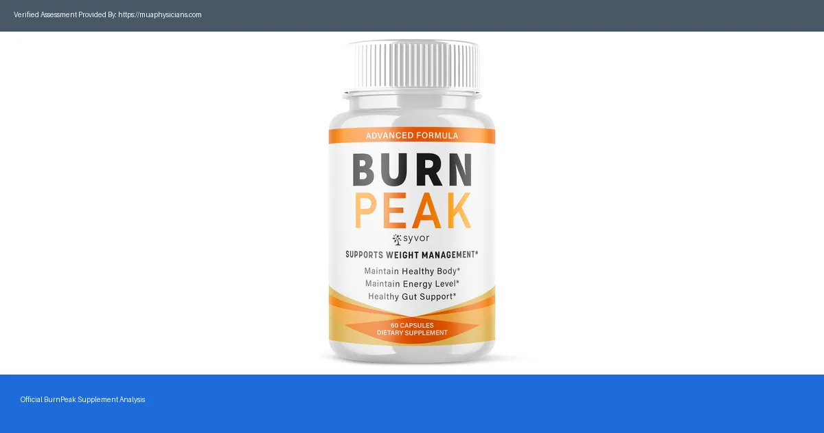
Discover the potential dangers of BurnPeak. Uncover surprising side effects and protect yourself from possible scams before purchasing.